The Incredible Journey: How Babies’ Heads Mold During Birth, Insights from a Groundbreaking MRI Study
Originally published: 2025-11-02
Unveiling the Study: A Peek Inside the Birth Canal
Childbirth is one of nature’s most remarkable processes, but much of what happens inside the body during labor has remained hidden, until now. In a pioneering 2019 study, researchers at the University Hospital Center in Clermont-Ferrand, France, used advanced MRI technology to capture 3D images of babies navigating the birth canal. This wasn’t just theoretical; seven brave women actually gave birth in an MRI machine, allowing scientists to document the dramatic transformations in fetal heads and brains during the second stage of labor. The goal was to understand fetal head molding, the way a baby’s skull compresses and reshapes to fit through the pelvis, and its effects on the brain.
CLICK HERE to review the study
“This study demonstrates the value of 3D MRI study with 3D finite element mesh reconstruction during the second stage of labor to reveal how the fetal brain is impacted by the molding of the cranial bones.” – From the study abstract
How It Was Done: The MRI Magic
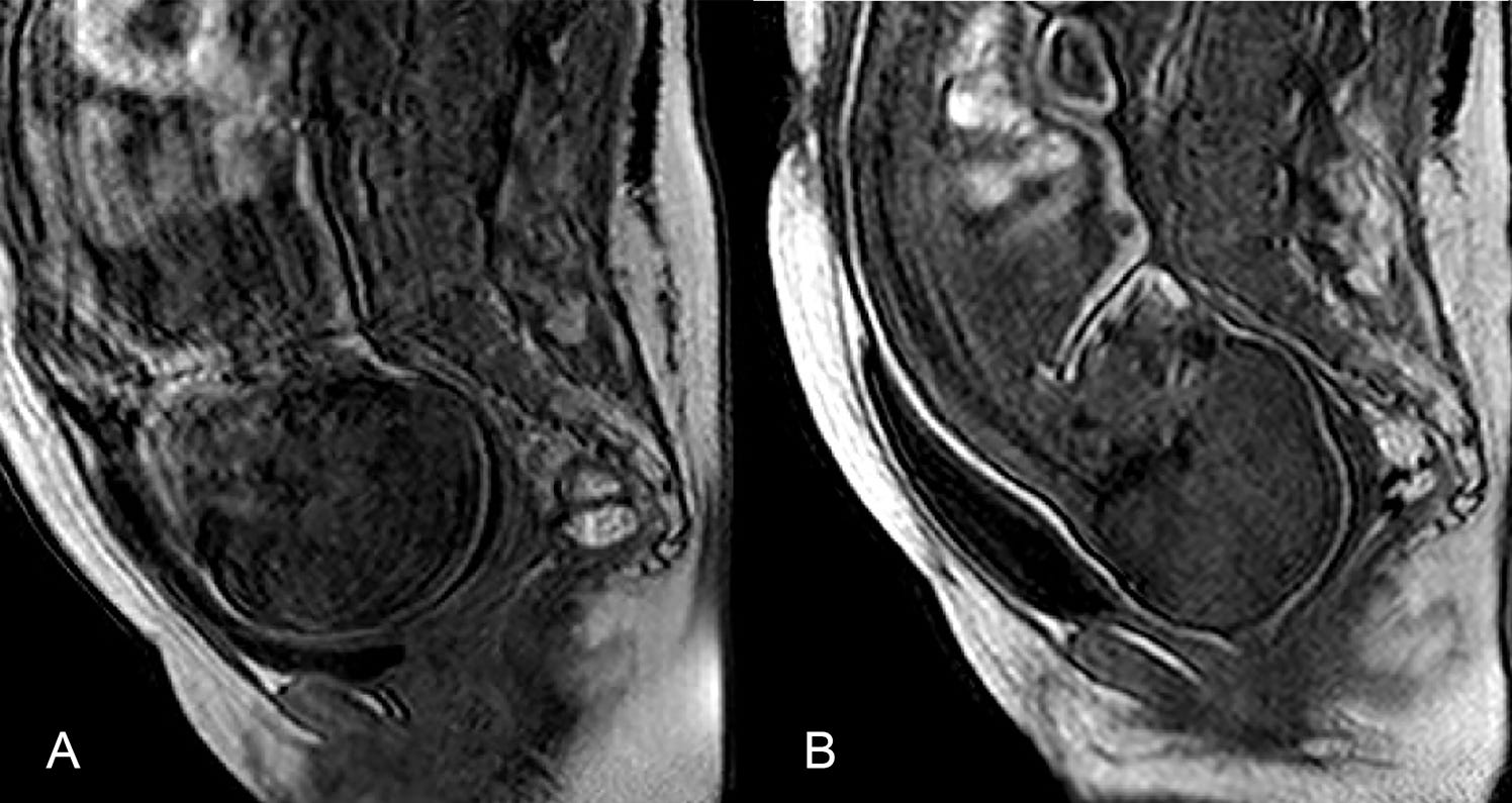
The research, titled “Three-dimensional magnetic resonance imaging of fetal head molding and brain shape changes during the second stage of labor,” involved 27 pregnant women initially scanned with 3D MRI between 36 and 39 weeks of gestation. Seven of them returned for scans during active labor, right after full cervical dilation but before pushing began. Using a 1 Tesla open-field MRI, the team captured fast, contiguous sequences without contrast, ensuring safety for both mother and baby. Images were then reconstructed into 3D models, aligning them to maternal pelvic landmarks for accurate comparisons.
Safety was paramount: participants had epidurals, were monitored by a full team, and could be rushed to the delivery room in under three minutes. The study focused on low-risk pregnancies, with women aged 23–34, and all signed informed consent.
What They Found: The Molding Miracle
The results were eye-opening. Every one of the seven fetuses showed varying degrees of head molding during labor, with skull bones overlapping, particularly at the coronal and lambdoid sutures, to create a “sugarloaf” shape that eases passage through the birth canal. The fronto-occipital diameter (front-to-back head length) decreased significantly, correlating with how far the baby had descended. Brain shape changed too, mirroring the skull’s compression, with fontanelles shrinking and others enlarging.
Post-birth, five of the seven babies’ heads snapped back to their pre-labor shape almost immediately, showing no lasting deformation on clinical exams. Images compared the fetus resting in utero (round and relaxed) to its compressed form in the canal, highlighting the skull’s pliable plates designed for this very purpose.
“When we showed the fetal head changing shape, we discovered that we had underestimated a lot of the brain compression during birth.” – Lead researcher Olivier Ami
The Impact on Brain and Birth Outcomes
Beyond the visuals, the study revealed potential stresses on the fetus. Greater molding was linked to two cesarean deliveries (one after failed forceps), suggesting that extreme deformation might signal complications. Interestingly, the baby with the most molding was born vaginally to a multiparous mother but showed brief distress, with a low initial Apgar score that quickly improved.
These findings hint at underestimated fetal stress, possibly explaining common post-birth issues like minor brain or retinal hemorrhages in vaginal deliveries. It also questions our definition of “normal” birth, emphasizing not just ease for the mother but the baby’s resilience to deformation.
Debunking Myths: No Such Thing as a “Too Big” Head?
Parents often hear that a baby’s head is “too big” for vaginal birth, leading to unnecessary interventions. But this study underscores that fetal skulls aren’t rigid, they’re built to mold. The overlapping plates and compressible brain allow most babies to navigate even tight spaces, with heads rebounding post-delivery. This challenges over-reliance on size predictions and highlights the body’s natural design for birth.
“Fetal head molding was systematically observed when the fetal head was engaged between the superior pelvic strait and the middle pelvic brim.” – From the study results
Final Thoughts: Celebrating the Spectacle of Birth
This French study isn’t just scientific, it’s a testament to the awe-inspiring adaptability of human bodies. By capturing these intimate moments, it empowers parents with knowledge, reduces fears around labor, and opens doors for future research into safer deliveries. Our babies and bodies truly are spectacular, as one birth advocate aptly put it. Next time you hear about a “big-headed” baby, remember, nature has a plan, and it’s more flexible than we think.
